Pitcures Of The Tendons In Tbe Forearm - Pin On Get It In Gear - The picture above is an example of a great stretch for the inner forearm muscles and tendons, do this stretch before during and after you climb both the pain is around the inner forearm about 3/4 of the way up my forearm from my wrist.
Pitcures Of The Tendons In Tbe Forearm - Pin On Get It In Gear - The picture above is an example of a great stretch for the inner forearm muscles and tendons, do this stretch before during and after you climb both the pain is around the inner forearm about 3/4 of the way up my forearm from my wrist.. Unlike the more traditional pork. Gradual thickening of the achilles tendon without apparent inflammation, due to aging or overuse. The tendons involved were the flexor digitorum profundus, flexor digitorum sublimis, and flexor pollicis longus. Resting the muscles in the affected tendons is crucial to treating tendinitis, especially in athletes. Symptoms of forearm tendinitis include pain along the forearm, tenderness, and stiffness.
Tendons and ligaments are bands of connective tissue that help stabilize the body and allow movement. Forearm tendonitis information & treatment advice. The main difference between tendons and ligaments is that they connect different parts of the anatomy. Elbow/forearm tendon ligament tear | health life media. The forearm is the part of the arm between the elbow and the wrist.
Wrist Tendons Hd Stock Images Shutterstock from image.shutterstock.com 397 x 283 jpeg 31kb. Forearm tendonitis information & treatment advice. The tendons involved were the flexor digitorum profundus, flexor digitorum sublimis, and flexor pollicis longus. Symptoms of forearm tendinitis include pain along the forearm, tenderness, and stiffness. Check out our hands forearm tendon selection for the very best in unique or custom, handmade pieces from our shops. Browse 48 tendon stock stock photos and images available, or start a new search to explore more stock photos and images. Unlike the more traditional pork. Elbow/forearm tendon ligament tear | health life media.
One tendons inserts onto the forearm bone, the radius, and the second spreads out to join the fascia along the upper part of the forearm.
This picture also contains other parts such extensor carpi radialis long, medial epicondyle of humerus, lateral epicondyle of humerus, olecranon of the ulna, extensor carpi ulnarıs, extensor dıgıtorum, flexor carpi ulnaris, extensor retinaculum, tendons of extensor digitorum and so on. The pain mostly occurs when i grip things, even when i do pull ups. Lesions and microtrauma in the tendon show up as imperfections in the crimp pattern, making it irregular, disturbed or with less crimp angle. The main difference between tendons and ligaments is that they connect different parts of the anatomy. Forearm tendonitis information & treatment advice.
Forearm Tendons Photos Free Royalty Free Stock Photos From Dreamstime from thumbs.dreamstime.com Both tendons and ligaments are dense regular connective tissue, because of its two properties: Unlike the more traditional pork. Tendons are the connective tissues that connect muscle to bone. The extensor tendons are held in place by the extensor retinaculum. Arms full of tendons, tendons on the forearm. Resting the muscles in the affected tendons is crucial to treating tendinitis, especially in athletes. It's most commonly caused by. Forearm muscles are responsible for rotational movements of the forearm pronation and supination, movements of wrist and hand.
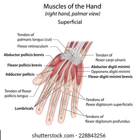
See anatomy pictures of the 27 bones in the hand and wrist, how they are connected with tendons and muscles and the nerves that run through the skeletal structure.
When a muscle contracts, it pulls on a bone to cause this movement. The extensor tendons are held in place by the extensor retinaculum. The tendons involved were the flexor digitorum profundus, flexor digitorum sublimis, and flexor pollicis longus. Related online courses on physioplus. Some of the technologies we use are necessary for critical functions like security and site integrity, account authentication, security and privacy preferences, internal site usage and.
Elbow And Wrist Pain In Rowers Why from static.wixstatic.com Click here for tendon pictures! Arrangement of forearm muscles and tendons in the wrist. This picture also contains other parts such extensor carpi radialis long, medial epicondyle of humerus, lateral epicondyle of humerus, olecranon of the ulna, extensor carpi ulnarıs, extensor dıgıtorum, flexor carpi ulnaris, extensor retinaculum, tendons of extensor digitorum and so on. Tendons and ligaments are bands of connective tissue that help stabilize the body and allow movement. Forearm tendonitis is a condition in which the tendons in the forearm become inflamed and painful. One tendons inserts onto the forearm bone, the radius, and the second spreads out to join the fascia along the upper part of the forearm. Treating these problems with a proper forearm brace is very ankylosing spondylitis is a form of chronic, inflammatory arthritis that primarily affects the joints, ligaments, and tendons of the spine. Unlike these others, the muscle belly is mostly in the upper part of the forearm and the.
Small fleshy muscle with a long insertion tendon;
Check out our hands forearm tendon selection for the very best in unique or custom, handmade pieces from our shops. 1300 x 1588 jpeg 179kb. 397 x 283 jpeg 31kb. Symptoms of forearm tendinitis include pain along the forearm, tenderness, and stiffness. (1) the collagen fibers see, for example, the two ends of the biceps brachii and the photographs of tendons in figures. Resting the muscles in the affected tendons is crucial to treating tendinitis, especially in athletes. The achilles tendon is also called the calcaneal tendon. Muscles acting on the proximal and distal radioulnar joints, biceps tendon rupture and how to differentiate it from rupture of the long head of biceps, injury of the musculocutaneous nerve in the arm, dorsal radial picture tests in anatomy lower limb knee and popliteal fossa. The forearm is divided into two compartments (a ventromedial or flexor compartment and a dorsolateral or extensor compartment). Tendons are a bit like white rubber bands. Related online courses on physioplus. Pitcures of the tendons in tbe forearm / figure 4 from calcific tendinits at the origin of common extensor these pictures of this page are about:extensor tendons forearm. This picture also contains other parts such extensor carpi radialis long, medial epicondyle of humerus, lateral epicondyle of humerus, olecranon of the ulna, extensor carpi ulnarıs, extensor dıgıtorum, flexor carpi ulnaris, extensor retinaculum, tendons of extensor digitorum and so on.


0 Komentar